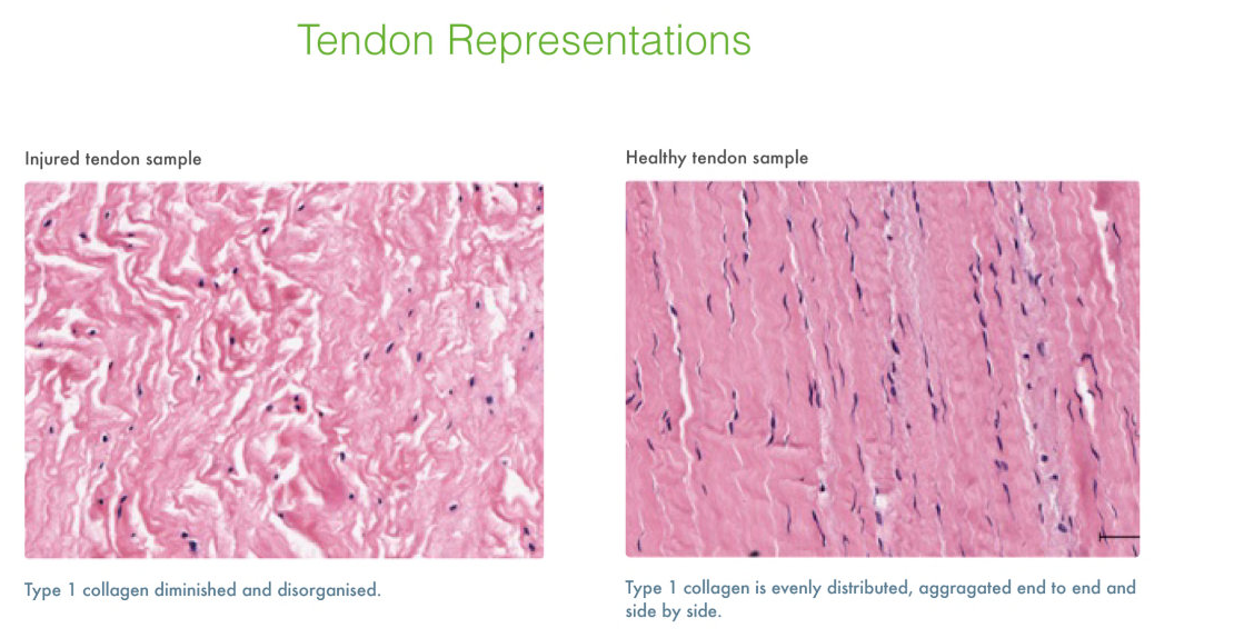
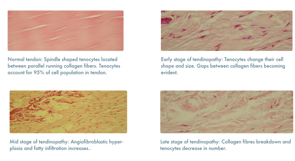
The pictures you posted are simply light microscopy of tissue (and prepared quite well). For a source, just look at the illustrations to Wikipedia's histopathology article and see for yourself what this type of picture looks like. Even if you could get a non-invasive imaging from a patient with that resolution (and I don't know of any which can do that, not even a 7 Tesla MRI), it won't look similar enough to be compared to the pictures from your post.
You'd need a biopsy of the tendon to get these images, and given how slow tendons are in healing, and that we are presumably dealing with an already damaged tendon, punching a piece out of it just to take a look is probably not a good clinical decision. Also, I don't know how easy or hard it is to get the tissue prepared in this quality in medical practice, I have seen such slides mostly in the context of research biology.
To make the answer complete: There are light microscope types which can be used in vivo, but they are certainly not yet ready for commercial use, not even in reasearch, much less in a clinical setting. And again, you're not really getting the same type of picture with them. So they are not a practical solution for what you want.

